WARP speed
Scientists from the University of St Andrews learn to measure the tiniest forces living cells exert at sheer WARP speed.
Writing in Nature Communications today (Friday 11 June 2021), the team of researchers, led by the School of Physics and Astronomy, have developed an advanced microscopy technique that greatly accelerates and simplifies our ability to study how cells interact with their environment.
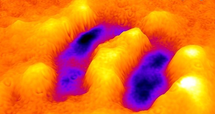
Wavelength alternating resonance pressure microscopy (WARP) provides near instantaneous maps of the extremely weak mechanical forces that living cells exert on their surroundings.
WARP allows scientists to record mechanical information about cells in real-time, a process that has previously required extensive offline computations to obtain results. Being able to record data in real-time opens a door to high throughput screening of the mechanical impact that new drugs may have on cells. It will also enable biologists who work with cell cultures to look at mechanical aspects more routinely than is currently possible.
The research team, led by Professor Malte Gather from the School of Physics and Astronomy at the University of St Andrews, demonstrated that WARP can resolve the forces that cause the periodic contractions of heart muscle cells. Of particular interest in this context is the mechanical characterisation of spontaneous oscillatory contraction (SPOC) waves, which are ripples of weak mechanical activity that flow through cells in between their main contractions.
SPOC waves may be involved in regulating the heartbeat by creating an intermediate phase between contraction and relaxation. The team are currently investigating using WARP to shed further light on this.
To measure mechanical forces, WARP employs thin film interference, the same phenomenon that gives soap-bubbles their rainbow-like colours. By illuminating a thin and soft probe with light of two slightly different colours in quick succession, the researchers can accurately and unambiguously infer by how much cells have deformed the probe.
The current work is part of a larger research effort funded by the Engineering and Physical Sciences Research Council (EPSRC) involving scientists from the University of St Andrews and the University of York that looks at exploiting optical resonances and the shaping of light to improve the understanding of biological systems.
Category Research