Laser cells offer ray of hope for understanding cancer
Researchers at the University of St Andrews have made a breakthrough in biomedicine; having successfully tracked a day-in-the-life of a number of white blood cells by feeding them microlasers, according to a research report published in Nano Letters today (Wednesday 22 July 2015).
The technique is expected to allow new insights into how cancers spread in the body.
The Soft Matter Photonics Group led by Professor Malte Gather of the School of Physics and Astronomy, in collaboration with immunologists in the University’s School of Medicine, found that by ‘swallowing’ an optical micro-resonator cells gain the ability to produce green laser light.
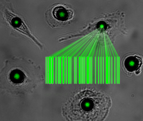
This miniature technology, which sounds like it might be more at home in the Ant Man movie, may soon develop into a useful tool in biomedical optics: the spectral composition of the laser light generated by each cell is different and can be used to distinguish and track large numbers of cells over prolonged periods of time. In their initial study the researchers demonstrate tagging of a few dozen human white blood cells and then track the cells for a day. In principle, the approach allows barcoding and reliably distinguishing up to several hundred thousand cells simultaneously.
Research groups around the world have worked on lasers based on single cells for several years now. However, all previously reported cell lasers required optical resonators that were much larger than the cell itself, meaning that the cell had to be inserted into these resonators. By drastically shrinking resonator size and exploiting the capability of cells to spontaneously take up foreign objects, the latest work now allows generation of laser light within a single living cell.
Dr Gather said:
“This miniaturization paves the way to applying cell lasers as a new tool in biophotonics. In the future, these new lasers can help us understand important processes in biomedicine. For instance, we may be able to track—one by one—a large number of cancer cells as they invade tissue or follow each immune cell migrating to a site of inflammation.”
Gather continued: “The ability to track the movement of large number of cells will widen our understanding of a number of important processes in biology. For instance being able to see where and when circulating tumour cells invade healthy tissue can provide insight into how cancers spread in the body which would allow scientists to develop more targeted therapies in the future.”
The investigators put different types of cells onto a diet of optical micro-resonators. Some types of cells were particularly quick to ‘swallow’ the resonators; macrophages—immune cells responsible amongst other things for ‘garbage collection’ in our body—internalized the resonators within less than five minutes. However, even cells without particularly pronounced capacity for endocytosis readily internalized the micro-resonators, showing that laser barcodes are applicable to many different cell types.
The micro-resonators used in this study were whispering gallery mode resonators. These are tiny plastic beads that trap light within a small volume by forcing it onto a circular path along the circumference of the bead. In the presence of an amplifying material, whispering gallery resonators emit laser light, even under relatively weak optical pumping conditions. This renders them ideal for integration into live cells as it prevents light-induced damage of the cells containing them. Indeed, in the present study, cells involved in the lasing process showed no loss in viability.
The spectral composition of the laser light emitted by whispering gallery resonators depends critically on the size and the refractive index of the plastic beads forming the resonator. Due to the inherent poly-dispersity (size variation) within a batch of whispering gallery resonators, a large number of unique lasing spectra are obtained.
Notes to news editors
The report is available to view online.
Artist’s impression of a group of cells which have been turned into tiny lasers is available from the Press Office on request.
About the Soft Matter Photonics Group:
The Soft Matter Photonics Group, which develops new photonic tools for the life sciences, was established at the School of Physics and Astronomy at the University of St Andrews in autumn 2013 with funds from the second phase of the Scottish University Physics Alliance and from other national and international funding bodies including the Human Frontier Science Program, the European Commission Seventh Framework Programme, and a H2020 Starting Grant from the European Research Council. The members of the group have diverse academic backgrounds ranging from physics to biology to chemistry. Professor Gather has previously led research groups at TU Dresden in Germany and worked at Harvard Medical School in the United States. Find out more about the Soft Matter Photonics Group.
Contact
University of St Andrews press Office on 01334 462 167 or email [email protected].
Category Research