Resolution revolution
Researchers at the University of St Andrews’ School of Physics and Astronomy have developed a new laser imaging technique that overcomes visual distortions and promises advances in deep tissue imaging and even nanosurgery.
Since the advent of the laser, the last 50 years have seen amazing advances in our use and understanding of light. However, light scatters quickly when passing through a turbulent object or medium – rendering any imaging or light focusing difficult. A good example is peering through mist or a typical bathroom window, which randomly scatters the incident light making it difficult to see what is on the other side.
Now Researchers at the School of Physics and Astronomy have developed a novel method to shape or “sculpt” the wavefront of light so that it reforms itself after passing through a turbulent medium.
Writing in the journal Nature Photonics, Tomas Cizmar, Michael Mazilu and Kishan Dholakia describe how they can place a fluorescent or strongly scattering object within a turbulent media and then employ a signal from this to provide aberration correction for the object that they wish to see. The correction to the light beam is introduced to the system via a liquid crystal micro-display display unit similar to those found in data projectors. Perfect focusing is an extremely important attribute in modern biophotonics systems and the researchers demonstrated the power of their technique in the case of optical trapping in a complicated scattering environment.
Tomas Cizmar said:
“Such methods are opening up a new window in bio-photonics sciences. These techniques are extremely powerful as they eliminate the obstructing barrier of biological samples given by their random structure, responsible for light scattering and wavefront degradation.”
Co-author Michael Mazilu said:
“The propagation of light still holds many surprises and new challenges in fundamental optics – who could ever have thought we could “see” through such aberrations?”
Kishan Dholakia said:
“This opens up exciting new applications: one can imagine one day using optical trapping to make measurements within blood vessels. In the shorter term we should be able to apply the same physics for probing inside artificial crystals, deep tissue imaging and even nanosurgery for cells embedded well within tissue. The possibilities are immense.”
The researchers are now advancing their technique with the Schools of Biology and Medicine for interdisciplinary applications.
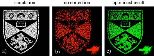
Optimization procedure applied on the crest logo of the University of St Andrews.
- Simulation of the resulting field distribution in an “optimal” system with no aberrations.
- The experimental reality obtained in our uncorrected system.
- Enhanced results after applying our correction method.
Issued by the University of St Andrews
Contact: Emma Shea, Communications Manager on 01334 462 109 or email [email protected].
Category Research